

Related products
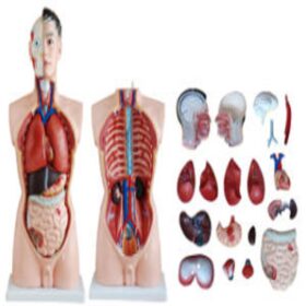


Half Body Human 15-Parts 26cm+brain | مجسم احشاء + دماغ
150,00 د.إ include VAT

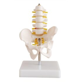

Pelvis With 5-Piece Lumbar Vertebrae Model | مجسم فقرات قطنية عجزية
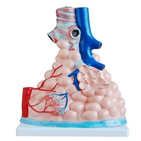
150,00 د.إ include VAT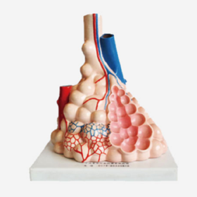 Magnified Pulmonary Alveoli Mo...
230,00 د.إ include VAT