Liver, Pancreas and Duodenum Model | مجسم تعليمي الكبد والبنكرياس
Brand:
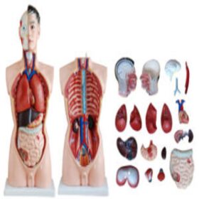
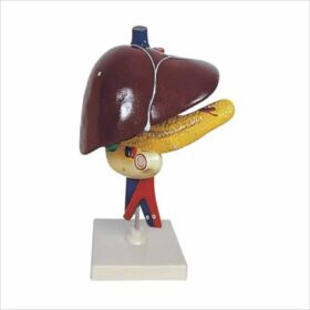
This model demonstrates the liver, spleen, blood vessels and pancreas. External structures are illustrated as well as the pancreatic duct of the pancreas. Also shows the abdominal aorta and inferior vena cava. Dissectible into 3 parts.
Anatomical Models of the Liver, Pancreas and Duodenum are essential educational tools used to demonstrate the complex spatial and functional relationships of the hepatobiliary and upper digestive systems. These models typically illustrate how the C-shaped duodenum curves around the head of the pancreas and receives bile and enzymes via a network of ducts.
Key Features of Professional Models
Life-Size Accuracy: Most high-quality models use a 1:1 scale to provide a realistic representation of human organ proportions.
Dissectible Parts: Many models are dissectible into 3 parts (liver, pancreas, and duodenum) or up to 6 parts (including the gallbladder, spleen, and stomach) to allow for internal inspection.
Vascular & Duct Systems: Models often highlight critical structures such as the pancreatic duct, common bile duct, abdominal aorta, inferior vena cava, and superior mesenteric vessels.
Materials: Constructed from durable, non-toxic PVC plastic, these models are hand-painted with realistic colors to distinguish different anatomical features.
Common Educational Uses
Classroom Instruction: Facilitates hands-on learning for anatomy and physiology students to understand the digestive process.
Clinical Demonstrations: Used by doctors and surgeons to explain conditions like gallstones, pancreatitis, or duodenal ulcers to patients.


 Liver, Pancreas and Duodenum M...