Sale

Related products


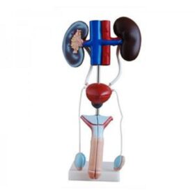
Male Genitourinary System | مجسم جهاز التناسلي الذكوري
150,00 د.إ include VATSale!
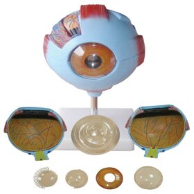

Giant Eye Model | مجسم عين
Original price was: 210,00 د.إ.175,00 د.إCurrent price is: 175,00 د.إ. include VAT مجسم حوض | Life-Size Hip Joint...
Original price was: 367,50 د.إ.300,00 د.إCurrent price is: 300,00 د.إ. include VAT