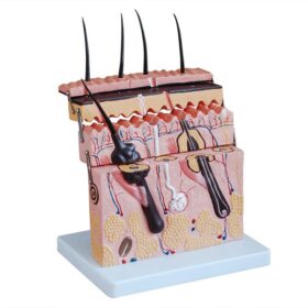
The Anatomical Skin Section Model is a detailed educational tool designed to showcase the multi-layered structure of human skin and its associated appendages. Typically constructed from durable PVC or HSP resin, this model is used in biology classrooms, medical training, and dermatology offices to visualize microscopic features like hair follicles and glands at an enlarged scale. Typically delivered mounted on a sturdy base (plastic or wooden) for easy display and examination.
- Dermatology: Used by specialists to explain skin conditions, disorders, or the depth of treatments.
- Medical Education: A standard tool for medical students to master the histology of the human scalp and other skin areas.
- Biology Classrooms: Ideal for high school and university levels to demonstrate how skin functions as a sensory organ and protective barrier.
- Three Primary Layers: Displays the epidermis, dermis, and hypodermis (subcutaneous fat layer).
- Skin Appendages: Features detailed representations of hair follicles (including the hair root), sebaceous glands, and sweat glands.
- Internal Systems: Shows intricate networks of blood vessels, nerves, and sense organs such as receptors.
- Hinged Design: Some versions are noted for having side hinges, allowing the model to be opened for a deeper view of the cross-section.


 مجسم مقطع جلد ثلاثي مع شعر | A...