

Related products


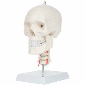

Skull With Cervical Spine Model | مجسم جمجمة مع فقرات رقبية
275,00 د.إ include VAT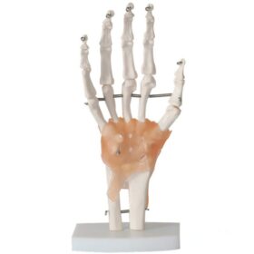
Life-Size Hand Joint with Ligaments | مجسم هيكل اليد مع الاربطة
150,00 د.إ include VAT Female Urogenital System Model...
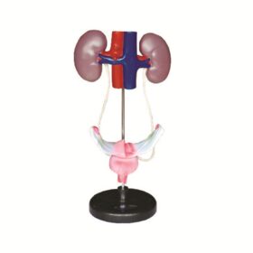
150,00 د.إ include VAT